
Beginning with the formation in week 9 and the development of the Muscular reflex in week 14 to the opening in weeks 22 thru 29.
WE R speaking about the windows of the world, yes, the human Eye, an organ that collects the images & keeps U in touch with reality. It is one of the 5 senses that lets us know of an objects existence. Ceaselessly gathering information: our eyes, ears, tongue, nose, & sense of touch provide a rich flow of signals to the brain, which in turn interprets them as reality. Aided by memory, for ex. The sound of a songbird & the smell of a flower can conjure up the pleasant images of a country garden. For some of us on the other hand, the sight of blood can make U faint or vomit.
Some say that sight and hearing R the most important senses. I feel that they’re all equally important, especially in the technologically advanced world we live in today. Sight, Hearing & Smell R concentrated in the Head. This comparatively exposed position is not without its risks, so most of the actual cells R well protected by the bony structure of the skull – which also protects the brain. Having this composition allows short fast, nervous connections between the sensors themselves & the controlling & analyzing brain.
In fact the sense organs R, in reality, elaborate & highly sensitive extensions of the central nervous system. All of the sensations detected by them trigger minute electrical impulses which travel along direct nervous pathways to the brain. Once there, they R coordinated & processed to give an ever changing mental image of the world around us, stimulating a wide variety of conscious & unconscious responses.
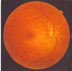
The structure of the eye: The area within the eye that detects light and color is called the Retina. The two types of detection cell present, rods and cones, process information coming through the Lens and send it down the optic nerve to the brain. Rod cells (of which there are around 100 million) detect the degree of light entering the eye, and their sensitivity is dependent on the amount of Rhodopsin present which is itself generated within the cells. However, Rhodopsin is destroyed by bleaching on exposure to light and therefore rod cells only work in low light as at high illumination the reduced level of this photosensitive pigment leads to a very low sensitivity. Cone cells (of which there are around 3 million) are also sensitive to light levels but retain their function up to high illumination via use of the pigment Iodopsin. Detection of color is a function of the three types of cone cells present within the retina: between them they cover the visible spectrum. This is because each type is sensitive to a different range of wavelengths with maximums corresponding to red (long), green (medium) or blue (short).
Our Eyes blink more than 10,000 times a day. & it accounts for approx. ¾ of our perception, using its 6 muscles to direct movement & anchor the eyeball deep inside bony bowls in the skull. With our eyes open, sensations come flooding in & R carried directly to the brain Via the optic nerve. When closed the tough fibrous plates of the eyelids form a waterproof & airtight shield over each eye. Sealing the eye opening from lid to lid, & lining the eyelids themselves, is the conjunctiva, a transparent membrane that catches anything that gets past the 1st line of defenses. It is kept moist by a thin layer of slightly oily tears produced by the lachrymal glands above the eye & spread by blinking. Tears contain a mild antibacterial agent, lysozyme, which provides the eyes with additional protection against harmful bacteria in the air. The sclera (white outer layer of the eye) has a transparent circular segment that bulges out at front to let light in; this is the cornea (bends incoming light directing toward the center). The exterior sclera’s appearance is opaque white, but its inner surface has a large # of blood vessels; this choroids layer is the main way which essential nutrients R supplied to the eye. The choroids also contains a layer of the dark pigment melanin, absorbing the right amount ensures that the interior of the eye remains dark, enabling the incoming light rays to give a perfect image on the retina. The cornea, a living tissue, in order to survive & function it has to have continuous supply of O2 & the proper Nutrients. Other body tissues obtain these vital requirements through the blood, but the cornea blood vessels would interfere with the passage of light. Therefore, it gets most of its O2 directly from the air, absorbing it through the tears. Nourishment comes via the aqueous humor, a watery transparent fluid that fills the space behind the cornea.
There r many types of genetic diseases 1 know as retinitis pigmentosa (RP). It degenerates peripheral vision. (It’s like tunnel vision) (The diagnosis of retinitis pigmentosa relies upon documentation of progressive loss in photoreceptor function by electroretinography (ERG){the patient assumes a comfortable position (lying down or sitting up). Usually the patient's eyes are dilated beforehand with standard dilating eye drops. Anesthetic drops are then placed in the eyes, causing them to become numb. The eyelids are then propped open with a speculum, and an electrode is gently placed on each eye with a device very similar to a contact lens. An additional electrode is placed on the skin to provide a ground for the very faint electrical signals produced by the retina. During an ERG recording session, the patient watches a standardized light stimulus, and the resulting signal is interpreted in terms of its amplitude (voltage) and time course. This test can even be performed in cooperative children, as well as sedated or anesthetized infants. The visual stimuli include flashes, called a flash ERG, and reversing checkerboard patterns, known as a pattern ERG}and visual field testing. The mode of inheritance of RP is determined by family history {Usher Syndrome [US] is inherited or passed from parents to their children through genes. Genes are located in every cell of the body and contain the instructions that tell cells what to do. Some genes specify traits such as hair color. Other genes are involved in the development of body parts, such as the ear. Still others determine how parts of the body work. Each person inherits two copies of each gene; one gene comes from each parent. Sometimes genes are altered or mutated. Mutated genes may cause cells to act differently, than expected. US is passed along in families by autosomal recessive inheritance, which requires two copies of the US gene before the disorder is seen. Each parent of a child with US usually has one standard and one mutated US gene. A child with US receives two mutated genes, one from each parent. Usually parents are unaware that they have or carry a US gene. This is because they would need two of the mutated genes in order to have signs of US. Presently, at least eight different genes are thought to cause the various types of US}. At least 35 different genes or loci are known to cause "nonsyndromic RP" (RP that is not the result of another disease or part of a wider syndrome). additional info: http://www.medicinenet.com/electroretinography/article.htm
Affected individuals first experience defective dark adaptation or nyctalopia (night blindness), followed by reduction of the peripheral visual field (known as tunnel vision) and, sometimes, loss of central vision late in the course of the disease.
There is great debate on the difference between wet / dry macula degeneration, new treatments R always on the horizon. Here is what I am able to explain.
Macula or Macula lutea (from Latin macula, "spot" + lutea, "yellow")
Chronic & Progressive, AMD (aged macular degeneration) is described as either wet or dry. Approx. 90% of patients have dry AMD, which typically progresses more slowly than the wet type. The other % of people has the wet AMD that probably started as dry AMD & then progressed.
1 of the most common early signs of dry AMD is the whitish yellow deposits called drusen, which develop under the retina. (Drusen alone doesn’t usually cause vision loss) an increased amount increases the risk of developing advanced wet/dry AMD.
Try to cut down on Sugar, white bread & other high – glycemic foods that spike blood sugar; it may promote this cause of blindness in older people.
Wet AMD aka Choroidal neovascularization (CNV) is also characterized by enlarged drusen. Weak abnormal blood vessels form beneath the retinal layers, leak blood & serous fluid into the macula, which is responsible for central vision. A damaged macula causes distorted vision & blind spots. The wet type is difficult to keep under control. In order to diagnose AMD, an ophthalmic specialist completes a comprehensive eye exam, using the following diagnostic tools:
more on AMD http://www.jofe-smolyak.com/maculardegeneration.html
Uveitis (you-vee-EYE-tis) is an irritation and swelling of the middle layer of the coats of the eye. This layer is called the uvea.
Uveitis can occur in one eye or both eyes. Inflammation of the uvea may involve other parts of the eye, or any part of the eye, including the cornea (the clear, curved front of the eye), the sclera (the white outer part of the eye), the vitreous body, the retina and the optic nerve.
Uvetitis may develop suddenly. U might feel pain in Ur eye or not & it may become red (inflamed) all over, Ur vision suddenly becomes blurry. Other symptoms of Uvetitis include light sensitivity, seeing spots. There r different kinds of Uveitis.
1) When the front portion of the uvea is swollen or inflamed (iritis)
2) When the middle layer is inflamed (cyclitis)
3) When there’s an inflammation of the back part of the uvea (choroiditis)
more on this topic http://www.preventblindness.org/uveitis/what/types.html
A thorough eye examination is necessary to diagnose uveitis. Ur ophthalmologist may order blood & skin, tests, or x-rays to determine whether U have uveitis or another condition. (uveitis can be caused by illnesses in other parts of the body). Some causes include having a fungus or a parasite infection. Uveitis may also be caused by an illness in another part of the body such as arthritis or lupus. Eye injuries, surgeries, can also lead to uveitis.
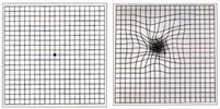
o An Amsler grid. The patient wears their daily glasses, covers 1 eye, holds the grid circa 14” (35mm) from the other eye, & looks at a large dot in the center of the grid. They should tell the ophthalmic specialist if any lines R wavy, bent, missing, discolored, blurry, if any of the boxes R a different shape or size.
http://www.amd.org/living-with-amd/resources-and-tools/31-amsler-grid.html
The patient wears their daily glasses, covers 1 eye, holds the grid circa 14” (35mm) from the other eye, & looks at a large dot in the center of the grid. They should tell the ophthalmic specialist if any lines R wavy, bent, missing, discolored, blurry, if any of the boxes R a different shape or size.
An
An Other common cause of blindness occurs when the pressure in the eyes rises, explains Dr. Tedd Mitchell in an article published by the USA weekend magazine, ----- damaging 1 of its most sensitive structures, the optic nerve. The disorders that cause this are grouped under the name Glaucoma.
The eye produces fluid to keep the chamber pressurized. In order to prevent the pressure from getting too high, the eye relies on “release valves” (1 way exits for excess fluid to escape and maintain a proper balance of pressure). Glaucoma occurs when the pressure created by the fluid in the eye rises, exceeding the amount necessary for appropriate function. Several different forms of glaucoma can cause this pressure imbalance to occur, but all create the same damage to the optic nerve if untreated. Anyone can develop glaucoma, but some groups are at higher risk: African Americans over 30, anyone over 60 & anyone with a family history of glaucoma.
An intake of high doses of antioxidant vitamins, including Zinc, Lutein, Lycopene, Bilberry,, Hawthorn Berries, may slow its progression, in addition to taking the antioxidants, exercise, quit smoking, maintain a healthy weight, control your blood pressure (see articles) & eat a healthy diet rich in leafy green vegetables & fish
(keeping the acidity levels in the body to a normal level; read Alkaline Vs Acidic article).
With Just 1 serving a month of kale or collard greens or more than 2 servings of carrots a week reduced the risk of glaucoma by more than 60% in a UCLA study of 1,000 women. Scientists believe that high levels of vitamin A & other antioxidants in these veggies help protect crucial cells in the optic nerve.
People who regularly drink orange juice & include C – rich foods like tomatoes, broccoli, red/green bell peppers in the daily diet R 45% less likely to develop cataracts. Vitamin C may help counteract the lens – clouding effects of light & heat.
Eating oatmeal, high – fiber cereals, & whole – grain breads cuts the risk of macular degeneration by about 39%, complex carbs prevent blood sugar swings that can damage delicate cells in the center of the retina.
Another symptom of blur can be caused by dry eyes here is a web-site with a detailed explanation http://www.eyecareamerica.org/eyecare/conditions/dry-eye/video.cfm
Eye dominance is an important consideration for monovision correction to reduce the need for reading glasses or bifocals. Anyone who is presbyopic should consider monovision. A test for eye dominance is with the both eyes open, raise your right arm and point to an object in the distance. Anything more than about 20 feet away is ideal. Continue to look at and point at the object and cover the left eye. Did your pointing finger seem to move off the target? Continue pointing and cover your right eye. Did your pointing finger seem to move off the target with your right eye covered? Your dominant eye is the eye you using when the pointing finger does not seem to move. Being right or left handed will not necessarily determine if you are right or left eye dominant.
The vitreous is the transparent, colorless, gelatinous mass that fills the space between the lens of the eye and the retina lining the back of the eye. It is produced by certain retinal cells. It contains very few cells (mostly phagocytes which remove unwanted cellular debris in the visual field, as well as the hyalocytes of Balazs, which reprocess the hyaluronic acid), no blood vessels, and 99% of its volume is water with salts, sugars, vitrosin, and a network of collagen type II fibers with the mucopolysaccharide hyaluronic acid accounting for the rest. The water content of the vitreous (98%) is greater than that of the lens (75%). However, the vitreous has a viscosity two to four times that of pure water, giving it a gelatinous consistency. It also has a refractive index of 1.336. Although the vitreous is in contact with the retina and helps to keep it in place by pressing it against the choroid, it does not adhere to the retina, except in three places: around the anterior border of the retina; in the macula, the tiny spot in the retina which gives us our "detail" and central vision; and at the optic nerve disc (where the retina sends about 1.2 million nerve fibres (axons) to the brain).
Unlike the fluid in the frontal parts of the eye (aqueous humour) which is continuously replenished, the gel in the vitreous chamber is stagnant. Therefore, if blood, cells or other byproducts of inflammation get into the vitreous, they will remain there unless removed surgically (floaters). If the vitreous pulls away from the retina, it is known as a vitreous detachment. As we age, the vitreous often liquefies and may collapse. This is more likely to occur, and occurs much earlier, in eyes that are nearsighted (myopia). It can also occur after injuries to the eye or inflammation in the eye (uveitis).
The collagen fibres of the vitreous are held apart by electrical charges. With ageing, these charges tend to reduce, and the fibres may clump together. Similarly, the gel may liquefy, a condition known as syneresis, allowing cells and other organic clusters to float freely within the vitreous humour. These allow floaters which are perceived in the visual field as spots or fibrous strands. Floaters are generally harmless, but the sudden onset of recurring floaters may signify a posterior vitreous detachment (PVD) or other diseases of the eye.
History has it that bilberry was 1st taken by British Royal Air Force pilots in WWII so they could see better on their nighttime bombing runs. Bilberry boosts Ur night vision, providing quicker adjustments to darkness & allows faster recovery from glare. In 1 study... documents that bilberry improves night vision by speeding up the filling of Visual purple (Rhodopsin) in the eye which enables better vision after dark, Bilberry helps improve:
☻ Eyes adjust quicker to darkness
☻ Allows Faster recovery after exposure to glare
Possible side effect: sensitivity to sun light.
Bilberry is good for people with chronic visual fatigue, including Pilots, Air-Traffic Controllers, Truck & Taxi Drivers, Computer Operators, Watchmakers, Researchers, & Students.
Improved night vision means greater safety, mobility & freedom.
Another Nutrient that is important is the Zeaxanthin. An Antioxidant found in fruits & vegetables such as Corn, Peaches, & Peppers. Zeaxanthin is vital for clear vision it protects the blood vessels & capillaries that supply the macular region: macula, retina, lens etc.
Zeaxanthin is one of the two carotenoids contained within the retina of the eye.
Within the central macula, zeaxanthin is the dominant component, whereas in the peripheral retina, lutein predominates.
Lutein and zeaxanthin have identical chemical formulas and are isomers, but they are not stereoisomers. The main difference between them is in the location of a double bond in one of the end rings. This difference gives lutein three chiral centers whereas zeaxanthin has two.
As a food additive, zeaxanthin is a food dye with E number E161h.
According to the Complete Guide to Vitamin & Minerals. “Dry eyes can occur as a result of Vit. A deficiency” Vitamin A deficiency can cause xerophthalmia/Xerosis (dryness of the conjunctiva), with the development of characteristic triangular or oval spots, called Bitot's spots.
Sun Damage can lead to cataracts, macular degeneration, & vision loss. Wear appropriated gear
|
1. Night Blindness |
Difficulty seeing in the dark. |
|
2. Xerosis (Dry Eyes) |
The white of the eye loses its shine and begins to wrinkle. |
|
3. Bitot's Spots |
Patches of little gray bubbles on the whites of the eye. |
|
4. Corneal Ulceration |
Dullness or damage to the cornea. |
|
5. Keratomalcia |
Soft or bulging cornea. |
Vitamin A is vital for the eyes health do to its maintenance of the integrity of membranes that circumferences the eyes that keep corneas moistened & lubricated. Vit. A is known to protect the eyes from inflammation.
See your health care provider if you have dry eyes. It may be a symptom of a more serious condition, such as rheumatoid arthritis or lupus. Also, constant irritation to the eye as a result of dryness can result in injury and damage.
Retinol also helps take care of an eye condition known as keratomalcia, (dry cornea)
People who depend on their eyes a lot knows the feeling of tired eyes, this is where taurine may come in handy Taurine (an amino acid) functions in active tissues such as the brain and heart to help stabilize cell membranes. It also has functions in the gallbladder, eyes, and blood vessels and appears to have some antioxidant and detoxifying activity. Taurine aids the movement of potassium, sodium, calcium, and magnesium in and out of cells and thus helps generate nerve impulses. Zinc seems to support this effect of taurine. Taurine is found in the central nervous system, skeletal muscle, and heart; it is very concentrated in the brain and high in the heart tissues, it revitalizes the eyes the natural way nourishing & stimulating the rods & cones of the retinas.
Alpha lipoic acid is a fatty acid found naturally inside every cell in the body. It's needed by the body to produce the energy for our body's normal functions. Alpha lipoic acid is also an antioxidant, a substance that neutralizes potentially harmful chemicals called free radicals. What makes alpha lipoic acid unique is that it functions in water and fat, unlike the more common antioxidants vitamins C and E, and it appears to be able to recycle antioxidants such as vitamin C and glutathione after they have been used up. Glutathione is an important antioxidant that helps the body eliminate potentially harmful substances. Alpha lipoic acid increases the formation of glutathione.
Alpha lipoic acid is made by the body and can be found in very small amounts in foods such as spinach, broccoli, peas, Brewer's yeast, brussel sprouts, rice bran, and organ meats. Alpha lipoic acid supplements are available in capsule form at health food stores, some drugstores, and online. For maximum absorption, the supplements should be taken on an empty stomach.
Alpha lipoic acid converts glucose (blood sugar) into energy Alpha lipoic acid helps neutralize a wide range of cell – damaging free radicals in the eyes & can penetrate eye tissue, protecting both the eyes` lenses & retinas, it has been suggested as a dietary supplement to treat diseases associated with excessive oxidant stress.
Another herb that could be helpful is Maritime Pine tree that grows along the Atlantic coast of the Bordeaux region of France this extract helps to control chronic venous insufficiency in several ways. 1st, chemicals called proanthocyanidins (or polyphenols) in the pine extract help keep veins and other blood vessels from leaking. A subgroup of proanthocyanidins is known as flavonoids. Catechin and taxifolin, two flavonoids in pine bark extract, may help blood vessels relax by increasing the production of nitric oxide, a natural body substance that helps keep blood vessels from becoming stiff and narrow. The extract has anti-inflammatory effects. Inflammation is often a response to irritation, injury, or infection and it usually includes pain, redness, and swelling in the area of the damage. Inflammation, which can occur within body tissues as well as on the surface of the skin, contributes to chronic venous insufficiency. The extract may also reduce the stickiness of blood components called platelets. Created in the bone marrow, platelets circulate in the blood. In a process called "platelet aggregation", they stick to injured tissue, beginning the blood clotting process and promoting wound healing.
Eye Chart Vision Test
A Snellen eye chart is used to determine how "normal" your vision is. It sets a standard for what most people should be able to see when they stand 20 feet away from the chart. 20/20 vision just means that when you stand 20 feet away from a Snellen eye chart, you see what a normal human being can see. If you see 20/40, that means that when you stand 20 feet away from the chart, you see what a normal person sees standing 40 feet away from it. The higher the second number, the worse your vision is. 20/200 (you see at 20 feet what a normal person sees at 200) is the number for legal blindness in the United States.
20/20 vision isn't perfect, it's just "normal." You can have better vision than 20/20. If you have 20/10 you see at 20 feet what most people see at 10. Some animals, like hawks, might have 20/2 vision!
You can use the Snellen eye chart*Click on the name for a printable version (The chart and the letters are named for a 19th-century Dutch ophthalmologist Hermann Snellen (1834-1908) who came up with them as a test of visual acuity. Visual acuity refers to the clarity or clearness of the vision, a measure of how well a person sees. The word "acuity" comes from the Latin "acuitas" = sharpness)to compare vision within your family or with your friends. (This will only give you an approximate idea of your vision. Your optometrist has much more precise tools to find out exactly how well you can see.) Each line of the chart is labeled on the left side. The second to last line is 20/20.
Tape the eye chart to a wall, making sure it is in plenty of light. Stand twenty feet away from the chart and begin reading each line. Have a family member or friend watch to see that you are reading each letter correctly. The last line that you are able to read will give you an approximate idea of your vision. If you can read the very bottom line, your vision is 20/10! Now try covering one eye and just testing the other one. Is one eye better than the other?
Have your entire family try reading the chart. Do some of you have better vision than others? If you wear glasses, what is your vision with them on and what is it without them?
Imagine knitting a scarf… working on a puzzle… fishing at dusk… enjoying the internet… or playing golf whenever U want & not worrying about Ur vision! Imagine yourself on a luxurious cruise… driving cross country…or watching a beautiful sunset - & not once having unfocused or cloudy vision. I think that is peace of mind don’t U?
Your eyes R not programmed to deteriorate with age. They do not have expiry dates stamped on them. They do not wear out with use like a pair of shoes. Eyes without the proper nutrition can start to deteriorate by presbyopia (long – arm disease) holding reading material far away from your eyes to obtain a clearer focus. Oxidative stress (free radicals) causes Ur lenses to thicken over time.
●●●
Don’t let unhealthy eyes ruin your independence.
Unhealthy eyes can mean: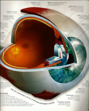
·
Failing Ur drivers, pilots, license renewal test…
·
Depending on others to take care of U…
·
Being “housebound” because of poor night vision…
· Declining invitations from family & friends….
·
Missing out on life’s great pleasures…
Eyeball has three layers
1. Fibrous tunic - this layer forms the sclera (opaque) and the cornea.
The Sclera is the white of the eye. It is avascular.
The Cornea is transparent and refractive. Also avscular.
2. Vascular tunic - the uvea. It consists of three parts:
a. Choroid - is highly vascular. It lines almost all of the internal surface of the sclera. It functions to reflect light back into the eyeball.
b. Ciliary body - forms a ring of fibers coming from ciliary muscles. These have ciliary process which attach to zonular fibers which in turn attach to the lens. The suspensory ligament of the lens is made up of these zonular fibers. When ciliary muscles are relaxed a constant tension is placed on the lens making it flatter. When ciliary muscles are contracted the tension on the lens is released and the lens becomes rounder.
c. Iris - is an extension of the choroid and is colored. It contains an opening called the pupil for regulation of light entering the eyeball proper. Constriction (contraction) of circular fibers (sphincter pupillae) results in a smaller pupil. Constriction of radial fibers (dilator pupillae) results in a larger pupil.
d. Lens - is made up of a transparent protein. It is highly refractive and its shape determines the amount of refraction.
A flat lens is better for viewing distant objects.
A round lens is better for viewing nearer objects.
3. Nervous tunic (Retina) - is generally divided onto an outer pigmented layer and an inner nervous layer. The inner nervous layer covers the posterior and lateral portions of the inner eyeball. Along the lateral margins (near the ciliary body) it thins to form the pigmented layer which covers the back of the ciliary body and iris
Chambers of the eye - the eye is divided into anterior and posterior portions.
1. Anterior cavity - is the portion anterior to the lens. It is subdivided into 2 chambers.
a. anterior chamber - located between the cornea and the iris.
b. posterior chamber - is located between the iris and the suspensory ligament of the lens.
Both of these chambers are filled with aqueous humor. It is a fluid which serves to maintain an interoccular pressure of about 24mm Hg. It also provides nutrients to the lens and cornea. This fluid drains via the canal of Schlemm, at the junction of the iris and cornea, into the bloodstream.
2. Vitreous chamber - is located between the lens and the retina. This chamber contains vitreous body, not humor. The vitreous body helps to hold the retina and the lens in place. It is not continually produced therefore if it is lost or damaged it is not replaced or repaired.
You should see an ophthalmologist or optometrist as soon as you notice any difficulty seeing, so you can get glasses or contact lenses. A sudden, radical change in vision may mean a serious medical condition. You should deal with it immediately. If you have young children, have their eyes checked before they start school and on a regular basis thereafter to prevent any trouble arising from limited or poor vision. If you are diabetic or at hereditary risk for glaucoma or other disorders, an ophthalmologist should monitor your condition on a regular basis
Brought to U by Nancy's Universe
Source of reference: Journal of Natural Alternatives Vol. 1 Issue 3, Wikipedia, http://www.healthy.net/scr/article.asp?ID=1971, http://en.wikipedia.org/wiki/Retinitis_pigmentosa, http://www.jwen.com/rp/rp.html, http://www.moondragon.org/health/disorders/xerophthalmia.html, http://www.drugdigest.org/DD/DVH/HerbsWho/0,3923,551937%7CFrench%2BMaritime%2BPine%2BBark%2BExtract,00.html, USA WEEKEND Jan 9-11 09, Readers Digest pg. 74 2/09, http://www.eyecareamerica.org/eyecare/conditions/uveitis/index.cfm, http://www.usaeyes.org/lasik/faq/lasik-monovision-dominant-eye.htm, http://www.hometrainingtools.com/articles/eye-chart-science-project.html,http://www.medterms.com/script/main/art.asp?articlekey=8552, http://en.wikipedia.org/wiki/Vitreous_humour, http://www.etsu.edu/cpah/hsci/forsman/WebVision.htm,